 Professor
 Associate Professor
Ultrasonic measurements of physical properties such like viscoelasticity of biological tissues and organs are investigated to realize their quantitative diagnosis, in addition to qualitative diagnosis based on conventional ultrasonic images. Researchers acquiring deep knowledge and excellent ability in engineering and fundamental knowledge in medicine, such like physiology, are nurtured through research and development of methods for controlling ultrasonic fields, ultrasonic measurements, and digital signal processing required for the quantitative measurements.
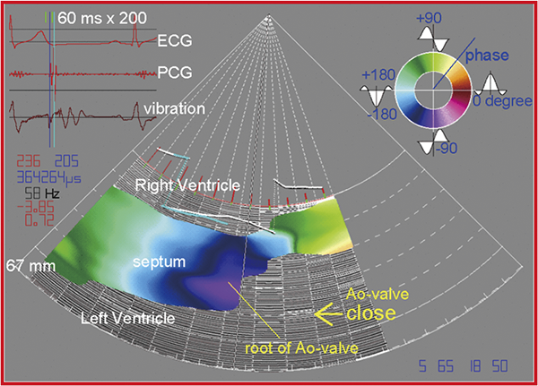
Imaging of propagation of spontaneous vibration in the heart wall
![Top: Microscopic image of normal (left) and aggregated (right) red blood cells (RBCs) ([1] J. R. Privitera et al., Silent clots: Life's biggest killers, Translated by K. Ujiie. Chuo Art, Tokyo.)
Bottom: Change in the sizes of ultrasonic scatterers (RBCs) due to aggregation during avascularization](../images/labo_01/labo_01_01-02.png)
Top: Microscopic image of normal (left) and aggregated (right) red blood cells (RBCs) ([1] J. R. Privitera et al., Silent clots: Life's biggest killers, Translated by K. Ujiie. Chuo Art, Tokyo.)
Bottom: Change in the sizes of ultrasonic scatterers (RBCs) due to aggregation during avascularization
 Professor
Sensing technologies are essential for the bio-electronic interface. For rapid and reliable analysis of biomolecules, highly sensitive and selective sensors are required for detection, measurement and visualization of specific molecules and ions. In this laboratory, chemical and biosensing technologies are developed based on semiconductor devices, which would be applied to biology and medicine.
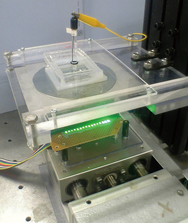
Chemical imaging sensor system
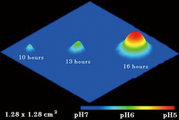
Visualization of pH by the chemical imaging sensor
 Associate Professor
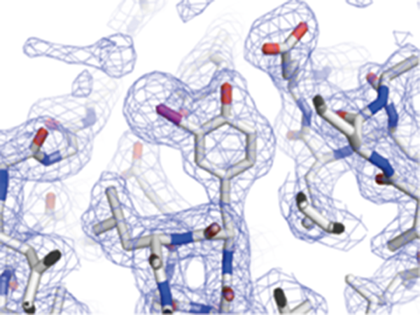
As gene products, proteins are concerned with many biological phenomena and they are key molecules to understand the mechanisms for diseases. A protein structure has close relationship with its function; therefore, revealing protein structures is very important for understanding protein functions. Although analyses of structural details for multidomain proteins or complexes are not simple task to achieve, the combination of various measurements (x-ray crystallography, molecular spectroscopies, mass spectrometry, etc) enables us to investigate mechanisms of diseases as well as to design new drugs.
Chemical imaging sensor system
Visualization of pH by the chemical imaging sensor
 Professor
Radiation is widely utilized for medical field as diagnostic and therapeutic tools. Radiation gives us information of living organism noninvasively. However, detected signals are in a tangle from several sources. We will investigate and develop advanced techniques to extract useful information from medical imaging including PET (positron emission tomography), SPECT (single photon emission computed tomography) and other modalities.
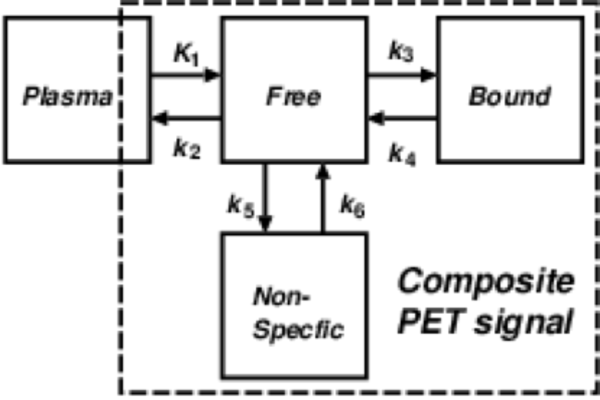
General mathematical model for analyzing PET data
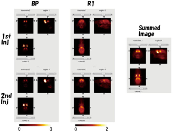
Generated parametric images from PET images with a rat