 Professor
Sensing technologies are essential for the bio-electronic interface. For rapid and reliable analysis of biomolecules, highly sensitive and selective sensors are required for detection, measurement and visualization of specific molecules and ions. In this laboratory, chemical and biosensing technologies are developed based on semiconductor devices, which would be applied to biology and medicine.
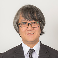
Chemical imaging sensor system
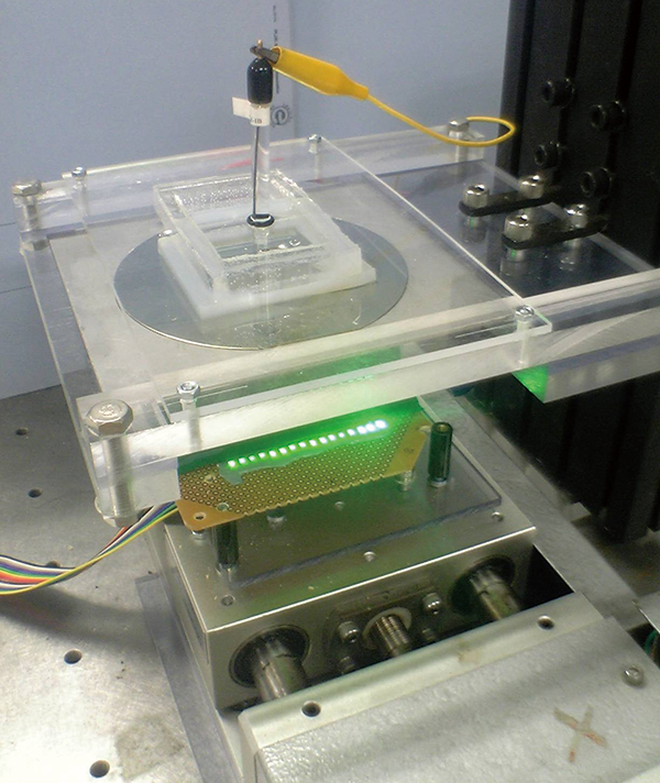
Visualization of pH by the chemical imaging sensor
 Associate Professor
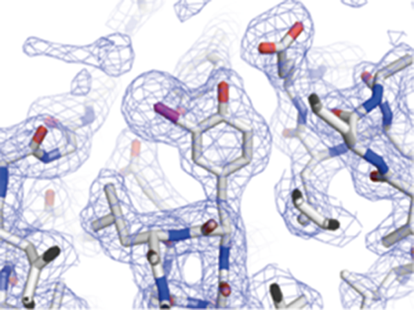
As gene products, proteins are concerned with many biological phenomena and they are key molecules to understand the mechanisms for diseases. A protein structure has close relationship with its function; therefore, revealing protein structures is very important for understanding protein functions. Although analyses of structural details for multidomain proteins or complexes are not simple task to achieve, the combination of various measurements (x-ray crystallography, molecular spectroscopies, mass spectrometry, etc) enables us to investigate mechanisms of diseases as well as to design new drugs.
Chemical imaging sensor system
Visualization of pH by the chemical imaging sensor
 Professor
Radiation is widely utilized for medical field as diagnostic and therapeutic tools. Radiation gives us information of living organism noninvasively. However, detected signals are in a tangle from several sources. We will investigate and develop advanced techniques to extract useful information from medical imaging including PET (positron emission tomography), SPECT (single photon emission computed tomography) and other modalities.
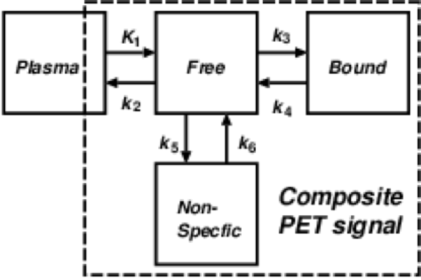
General mathematical model for analyzing PET data
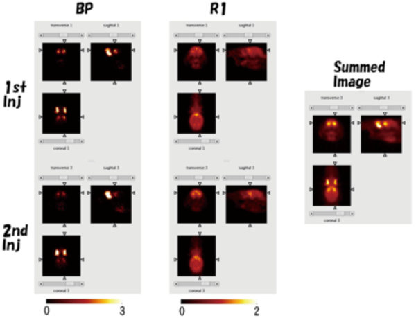
Generated parametric images from PET images with a rat
 Associate Professor
Our research is situated at the intersection of life sciences, medicine, and engineering, with a dedicated focus on unraveling the complexities of neurological and psychiatric disorders, which are major societal challenges. Our team specializes in the research and development of microelectronic fiber technology that enables simultaneous measurement and manipulation of diverse signals across in vitro, in vivo, and wearable settings. Leveraging a thermal drawing technique similar to the process used to make "Kintaro candy," our approach facilitates the integration of multiple functionalities—such as optical waveguides, electrodes, microfluidic channels, biosensors, and actuators—into a single, soft and flexible fiber. The multifunctional fibers we develop are instrumental not only in foundational research on neurological and psychiatric disorders but also in advancing new diagnostic and therapeutic tools in healthcare and medical treatments.
Our key research areas include:
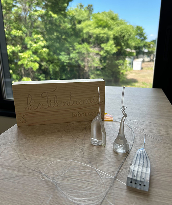
Microelectronic fibers
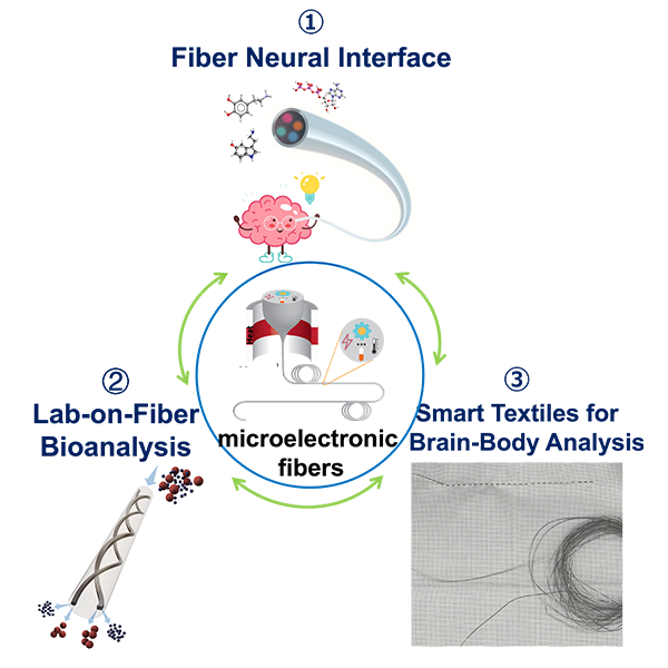
Research diagram
 Associate Professor
We are conducting Research and development of medical imaging technology (X-ray imaging, nuclear medicine imaging) based on the following two approaches. The first is the development of technologies that link hardware and software to maximize numerical performance indicators of imaging technologies such as resolution, sensitivity, and noise characteristics. In many cases, these have physical limitations, and the goal is to develop technologies that approach or exceed these limitations. Second, the goal is to provide technology that allows physicians, the users of the images, to safely perform medical procedures. Based on the measurement of biometric information (eye movements and cranial nerve activity) while a person is looking at a medical image, we aim to build technology to reduce the risk of medical accidents/errors.
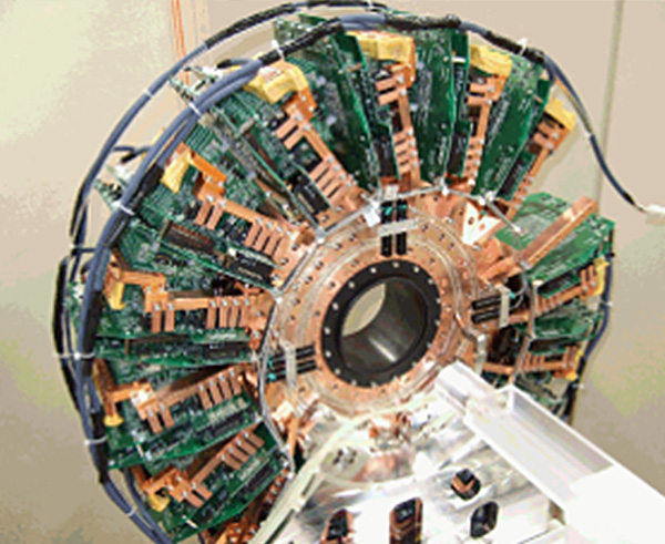
Development of super-resolution nuclear imaging
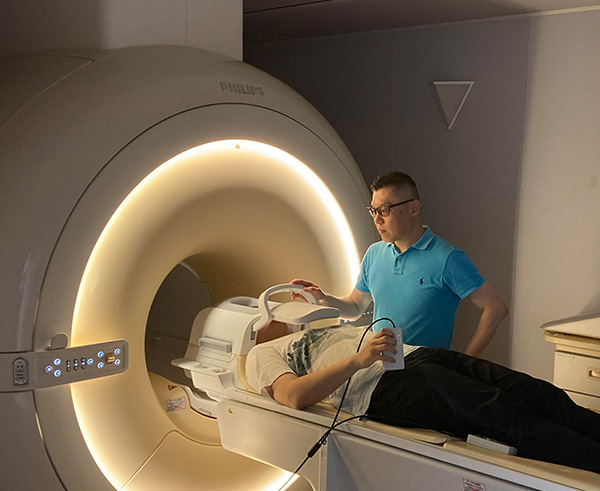
Evaluation of medical imagin based on brain activity measurement using MRI