 Professor Masayoshi Mizutani
 Our laboratory aims to promote innovations of nano-precision Micro/Meso Mechanical Manufacturing (M4 process) at the frontier of manufacturing technology for a smart functional interface. The geometric structure and chemical composition of material surfaces change in a variety of ways under machining conditions. Our laboratory examine this phenomenon and attempt to clarify and control the mechanism. This method can create various surface functions, such as biocompatibility, antibacterial activity, and wettability, which could lead to new surface creation processes. Our goal is to create new principle and technology for the next-future bio-medical interface and devices.
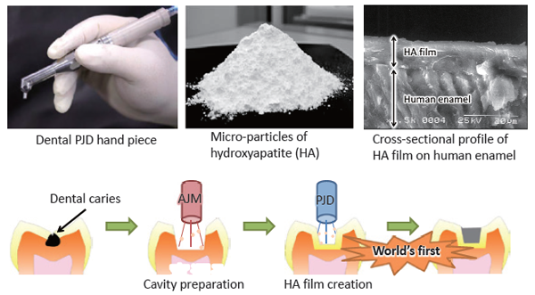
New dental treatment utilizing powder jet deposition of hydroxyapatite
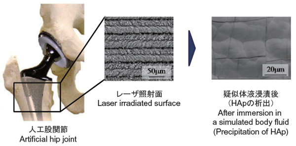
Generation of bio-active surface
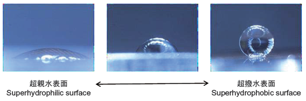
An example of surface function: surface wettability control
 Professor
Small and high-functioning and multi-functioning endoscopes, surgical tools have been developed using micro fabrication technologies, for example, micromachining, nanotechnology and MEMS (Micro Electro Mechanical Systems) technology aiming to realize practical and useful medical devices in near future, and we are aiming to realize robot surgery and microsurgery from inside the human body in the future. Microsensors, microactuators, and batch fabrication technologies for low cost and precise production have been studied and developed to realize the above. Utilizing these microfabrication technologies, we also aim for healthcare applications to realize new widely useful measurement items and methods and to develop thin and light weight wearable healthcare devices. Furthermore, organ models, for example vascular model and brain model, equipped with micro sensors have been developed to contribute surgical training of medical doctors and to evaluate effectiveness and safety of developing new medical devices.
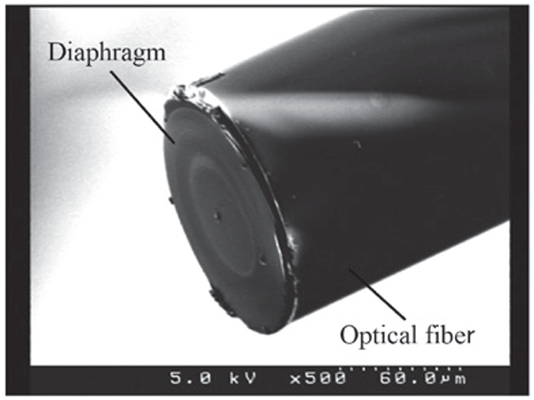
Ultra-miniature fiber-optic pressure sensor (O.D. 125µm)
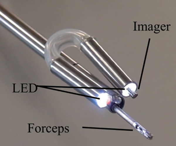
Bending Transformative Endoscope for Intraperitoneal surgery (O.D. 5mm)
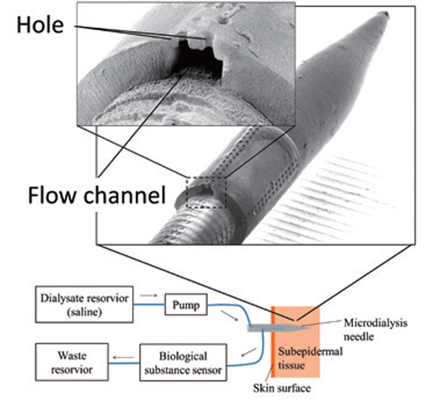
Metal needle with micro flow channel for biological substances monitoring patch Cross-section view of flow channel (needle O.D. 200µm).
 Professor
 Assistant Professor
Our laboratory is developing signal and image processing methods of ultrasound, CT (computed tomography) and MRI (magnetic resonance imaging) in order to realize three-dimensional imaging, precise automatic diagnosis and flow analysis of cardiovascular tissues. We are also developing several acoustic microscopes to visualize kidney, liver, tendon, cartilage, bone and tooth besides cardiovascular tissues and assessing pathophysiology from the view point of biomechanics.
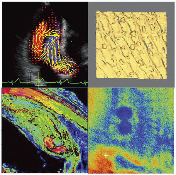
Upper left: Blood flow vector in left ventricle
Upper right: Three-dimensional ultrasound image of fingerprint
Lower left: Ultrasound microscope image of coronary artery
Lower right: Ultrasound microscope image of living cell
 Professor
We are conducting research on non-invasive and minimally invasive healthcare systems that utilize optical technologies such as infrared and ultraviolet light. These systems do not require a blood draw; they can analyze blood components simply by irradiating the body with light. Additionally, by simply breathing into an optical sensor, they can display health metrics to monitor your physical condition. By developing systems that utilize optical technologies, such as laser devices, to analyze various components of biological tissue, we aim to create systems that support daily health management and enable rapid, highly accurate disease diagnosis.
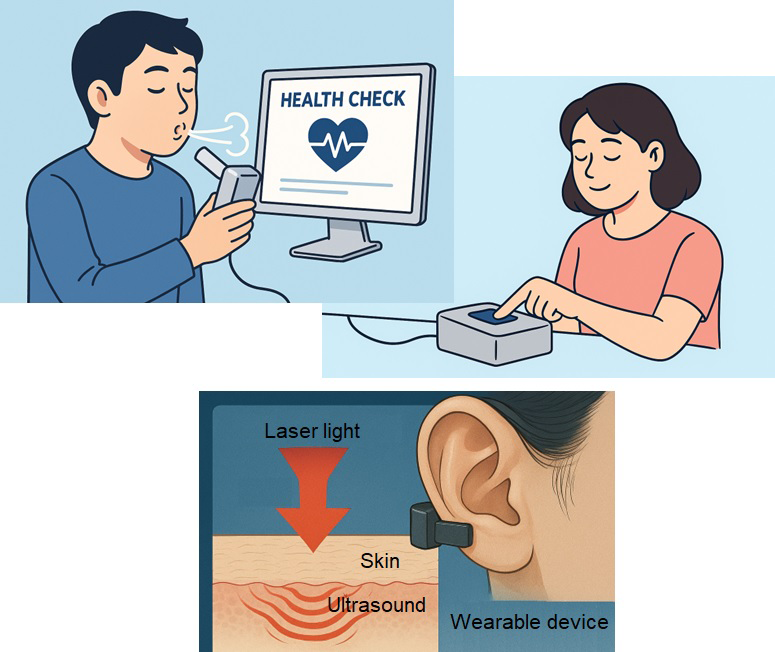
Just touch the sensor or blow on it for a health check. An image of a wearable sensor.
 Professor
 Associate Professor
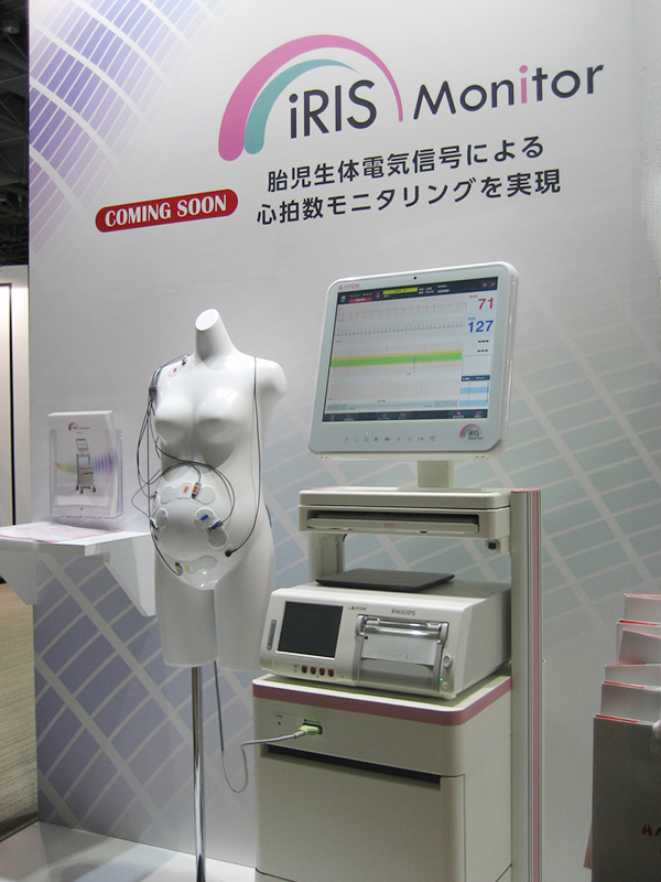
It has been found that some environments around fetus, such as maternal diet and infection, affect the baby’s growth or constitution significantly.
For example, the excessive intake of fat diet of mother during pregnancy is due to recent increase of children's autism, juvenile diabetes, and air pollution around mother has been found the cause of increase in childhood asthma.
In spite of the recent advanced technology, most of the precise mechanism of fetal differentiation process has been unknown in the black box named uterus.
In this laboratory, we study the fetal diseases from the relationship between maternal conditions and fetal development using the mice experiments, gene analysis, and clinical studies. And we explore the medical engineering in the near future though the study of measurement information obtained from the faint fetal signals appeared from maternal body.
 Professor
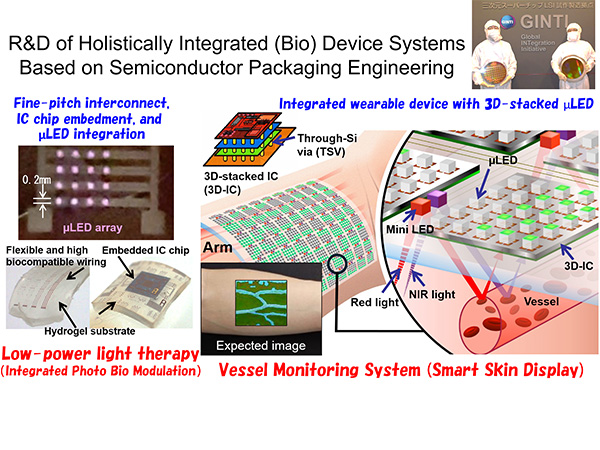
Holistic Integration Engineering is a research field that uses a systematic holistic approach to device creation, based on semiconductor packaging engineering, which significantly affects the performance of integrated circuits. We are researching manufacturing technology that contributes to cutting-edge AI chips and high-performance wearable devices through valuable experience using a cleanroom that is on a par with the best in the world.